Treatments

Narrow angle glaucoma laser treatmento – Yag laser iridotomy. * Narrow-angle glaucoma is a severe, acute form of the disease. In this type of glaucoma, the iris of the eye is pushed forward, blocking the channel (angle) that normally allows aqueous fluid to drain. The resulting spike in intraocular pressure causes an acute attack of glaucoma, which may permanently damage the optic nerve.
Laser peripheral iridotomy is the standard first-line treatment in closed angle glaucoma and eyes at risk for this condition. With this laser your eye doctor creates a hole in the outer edge of the iris, leading to an opening of the angle in the majority of cases. After the angle is widened from the procedure, the fluid outflow is enhanced and the intraocular pressure normalizes.
Retina laser treatment – laser Argon. * The retina is a delicate lining in the back of the eye where the images you see are made. Laser treatment involves using an intense beam of light (laser) that can be precisely focused to treat certain diseases of the retina. This treatment is done to make sure your current level of vision lasts and to prevent any further loss of vision. There are many eye conditions which can be treated with laser, for example: diabetic eye disease, macular oedema, retinal tears or holes and occlusions of retinal blood vessels.
LASER TREATMENT
Laser has particular use in ophthalmology because the eye can be used as an optical device. The transparency of the front part of the eye, the cornea, allows light such as LASER to reach almost all the tissues of the eye. A beam of light can reshape a cornea to improve its focus, create a channel within the eye to relieve the intraocular pressure of glaucoma or cauterize tiny hemorrhages. Lasers have become as essential to the practicing ophthalmologist as the scalpel, the magnifier and sterile gloves.
Secondary cataract laser treatment – Yag laser capsulotomy. * Secondary cataract: Although some people call it “secondary cataract,” it really is not a cataract. Once a cataract is removed, it does not come back.
During cataract surgery, your surgeon will remove the cloudy natural lens of your eye (cataract) and replace it with an intraocular lens. Much of the thin clear membrane that surrounds the natural lens (called the lens capsule) is left intact during surgery and the artificial lens usually is implanted within it. When the cataract is removed, your surgeon makes every attempt to maintain the integrity of the lens capsule, and normally your vision after cataract surgery should be very clear. However, in about 30 percent of patients, the posterior portion of the capsule becomes hazy some time during cataract surgery recovery or even months later, causing secondary cataract.
Fortunately, a YAG laser can treat posterior capsule opacity safely, effectively and painlessly. This procedure, known as YAG laser capsulotomy, often can be performed in your doctor’s office. The laser removes the hazy posterior capsule from your line of sight without making an incision or “touching” the eye. The procedure takes only a few minutes and is entirely painless. Most people can expect their vision to improve within a day.
Open angle glaucoma laser treatment – SLT Selective laser trabeculoplasty. * Open-angle glaucoma is characterized by a gradual decrease in aqueous fluid outflow despite an open anterior chamber. It is a chronic, silent thief of sight. Once the condition has passed beyond the point at which the patient can control it with eyedrops or pills, an ophthalmologist can perform laser treatment.
Selective Laser Trabeculoplasty is a form of laser surgery that is used to lower intraocular pressure in glaucoma. It is used when eye drop medications are not lowering the eye pressure enough or are causing significant side effects. It may sometimes be used as initial treatment in glaucoma. Laser energy is applied to the drainage tissue in the eye. This starts a chemical and biological change in the tissue that results in better drainage of fluid through the drain and out of the eye.
INTRAOCULAR INJECTION
( Lucentis, Eylea, Vabysmo )
Intravitreal drug delivery has become a popular method of treatment of many retinal diseases, commonly including macular degeneration, diabetic eye disease and retinal vein occlusions. An intravitreal injection is a shot of medicine into the eye. The inside of the eye is filled with a jelly-like fluid (vitreous). During this procedure, your eye doctor injects medicine into the vitreous, near the retina at the back of the eye. The medicine can treat certain eye problems and help protect your vision. Your vision may remain stable or improve after the procedure. You may need more than one injection.
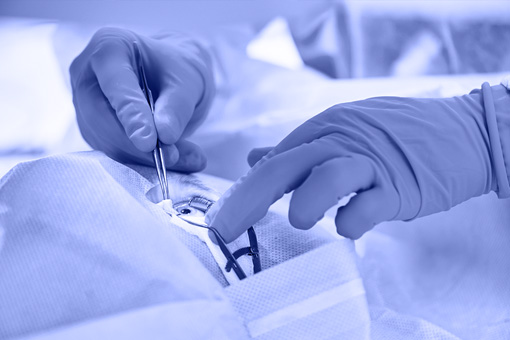
EYE SURGERY
Cataract surgery (phacoemulsification)
Modern cataract surgery is one of the safest and most effective surgical procedures performed today. In cataract surgery, the lens inside your eye that has become cloudy is removed and replaced with an artificial lens (called an intraocular lens) to restore clear vision. The procedure typically is performed on an outpatient basis and does not require an overnight stay in a hospital or other care facility. Most modern cataract procedures involve the use of a high-frequency ultrasound device that breaks up the cloudy lens into small pieces, which are then gently removed from the eye with suction. After all remnants of the cloudy lens have been removed from your eye, the cataract surgeon inserts a clear intraocular lens, positioning it securely behind the iris and pupil, in the same location your natural lens occupied.
Retinal surgery ( macula pucker, macula hole, retinal detachment)
For some cases of macula pucker or macula holes a surgery called vitrectomy is recommended. During surgery, your ophthalmologist uses microsurgery instruments to remove the wrinkled tissue on your macula and to remove the vitreous gel that may be pulling on the macula. Sometimes an air or gas bubble is placed in the eye to help the retina heal or to seal any tear or holes. After the tissue is gone, the macula flattens and vision slowly improves, though it usually does not return all the way to normal.
Retinal detachment repair is eye surgery to place a retina back into its normal position. The retina is the light-sensitive tissue in the back of the eye. Detachment means that it has pulled away from the layers of tissue around it. Most retinal detachment repair operations are urgent. A detached retina does not get a supply of oxygen. This causes the cells in the area to die, which can lead to blindness. The vitrectomy procedure uses very small devices inside the eye to release tension on the retina. This allows the retina to move back into its proper position.
Glaucoma surgery( trabeculectomy)
Glaucoma is a complicated disease in which damage to the optic nerve leads to progressive, irreversible vision loss. Currently the goal of glaucoma treatment is to reduce or stabilize intraocular pressure. When this goal is accomplished, damage to ocular structures, especially the optic nerve may be prevented. When medicines and laser surgeries do not lower eye pressure adequately, doctors may recommend a procedure called filtering surgery. Trabeculectomy is a minimally-invasive glaucoma procedure that increases flow into the natural drainage system of your eye. In filtering surgery, a tiny drainage hole is made in the sclera. The new drainage hole allows fluid to flow out of the eye and helps lower eye pressure. This prevents or reduces damage to the optic nerve.
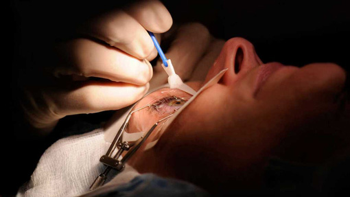
Eyelid surgery (eyelid growths, tumors, excess skin, blepharoplasty)
Surgery of eyelid can frequently be done under local anesthetic, with the surgeon assisted by magnification microscope in order to place very fine stitches which are almost invisible. The procedure normally takes aproximately 30 to 45 minutes.